
The intricate dance of pupil dilation and constriction can reveal invaluable insights into neurological function. Enter the Neurological Pupil Index (NPi), a pioneering metric that harnesses pupillary reactions to illuminate otherwise hidden signs of neurological conditions. Join us as we explore NPi’s revolutionary role in advancing the frontiers of neurological assessment.
Understanding the Neurological Pupil Index
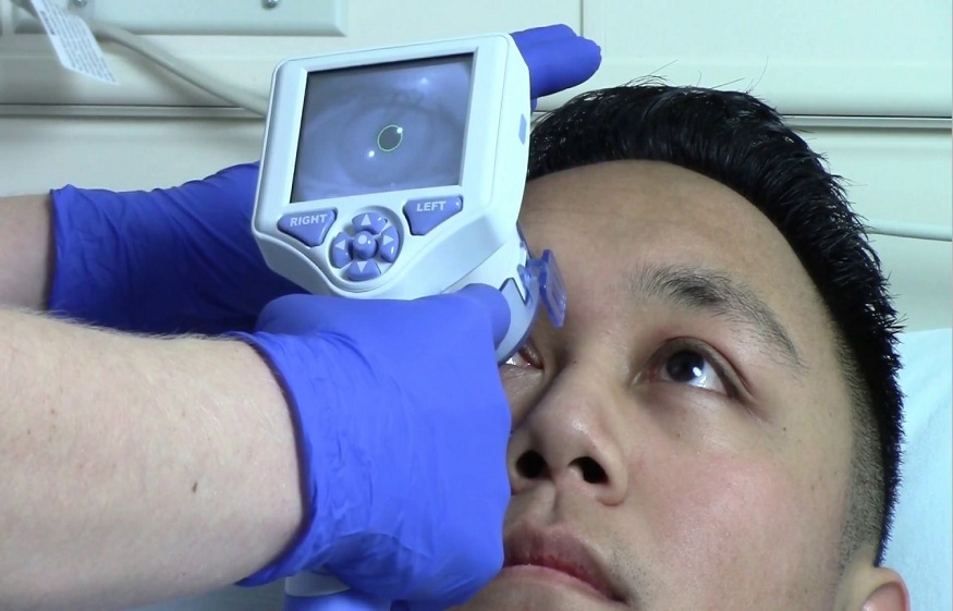
The Neurological Pupil Index (NPi) is a novel diagnostic tool that analyzes pupillary responses to shed light on neurological health. By tracking the percent change in pupil size, NPi provides an objective quantification of pupillary light reflexes. This gives neurologists an accurate and reliable way to uncover abnormalities that traditional penlight exams might miss.
NPi offers a whole new dimension to the neuro exam by providing quantitative data on pupillary function. Traditional methods like swinging a penlight and subjectively estimating pupillary reactions offer limited insight. NPi goes far beyond this by precisely measuring parameters like percentage change in pupil size, dilation and constriction velocity, diameter, and latency. This wealth of objective data opens up new possibilities for detecting neurological issues.
The Advantages of NPi in Neurodiagnostics
Unlike qualitative and subjective traditional pupil exams using inaccurate neurological tools, NPi offers quantitative pupil analysis. This allows for continuous objective monitoring of neurological function, which is perfect for prompt decision-making. NPi testing is also non-invasive, taking only seconds to perform. The real-time insight lets clinicians detect early neurological changes before deficits become debilitating.
NPi provides objective data that allows clinicians to precisely track pupillary function over time. This grants unique insights into disease progression, recovery, and treatment response. The scores are consistent across examiners, eliminating human errors and biases during traditional pupillary exams.
NPi’s Applications in Neurological Assessments
From concussion management to stroke evaluation, NPi is finding diverse clinical applications. For example, tracking NPi after head trauma provides objective data to guide return-to-play decisions. In stroke, falling NPi scores can indicate ischemia and precisely localize lesions. NPi also aids in tracking intracranial pressure changes, offering invaluable monitoring in cases of brain tumors or hemorrhages. Furthermore, NPi facilitates the prognosis of neurological recovery across diverse conditions.
By objectively quantifying pupillary abnormalities, NPi provides an early indicator of neurological disturbances in conditions like Alzheimer’s and multiple sclerosis. In concussion management, NPi is especially useful for objectively demonstrating recovery before a return to normal activity. Serial NPi measurements also aid in localizing lesions, monitoring edema and mass effects, and prognosticating stroke and brain injury outcomes.
NPi Paves the Way for Precision Neurology
NPi is paving the way for personalized neurological care. By creating objective pupillary profiles of individual patients, NPi enables tailored treatment plans. For instance, mild traumatic brain injury shows heterogeneity in pupillary deficits. NPi scores allow the subclassification of concussion phenotypes for targeted management. Likewise, NPi facilitates tracking patient-specific responses to neurological treatments. This aligns perfectly with the goals of precision medicine.
By moving beyond the “one-size-fits-all” approach, NPi provides the objective data needed to customize neurological treatment. The pupillary biomarkers captured by NPi offer insights into the unique pathophysiology underlying each patient’s condition. This opens doors for tailored therapeutic plans based on an individual’s pupillary phenotype.
Overcoming Current Limitations
While promising, NPi has boundaries. Testing conditions like ambient lighting can influence readings, underscoring the need for protocol standardization. Also, more research is needed to determine normative ranges across wider populations. Nonetheless, the future is bright for NPi. Ongoing studies on improving test protocols and developing pupil-tracking technologies will only strengthen NPi’s utility. With further validation, NPi may find routine application in diverse neurological disorders, from Parkinson’s to Alzheimer’s and beyond.
Factors like lighting, distance from the pupil, and recording angle can skew NPi results. This highlights the need to establish rigorous testing protocols. Extensive research is essential to defining normal NPi values across ages, ethnicities, and health statuses.
Conclusion:
The Neurological Pupil Index heralds an exciting frontier in neurodiagnostics. By decoding pupillary physiology, NPi provides objective insight into neurological function in an accessible and non-invasive manner. As NPi technologies and techniques continue maturing, this neurological “crystal ball” will likely transform the landscape of neurology and propel us toward personalized medicine for the nervous system.